![]()
UROGENITAL
SYSTEM
Urinary System
Avian kidneys lay in the abdomen of the bird near the back and sides (dorsolateral)
and excrete dilute uric acid that is later concentrated in the cloaca. The
ureters leave the kidneys from the middle of the 3-lobes and move waste to the
cloaca for concentration.
Birds secrete solid uric acid instead of liquid urine (urea) allowing for water
conservation, less weight to maximize flight ability and allowing for storage of
embryonic waste in the egg as a solid instead of liquid which would be toxic to
the developing chick. The kidneys also excrete salt and nitrogenous waste
products. The renal portal system is simple and can be shut down for short
periods during times of stress to allow for increased blood flow to other
organs.
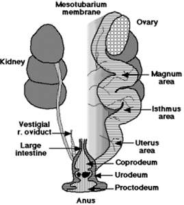
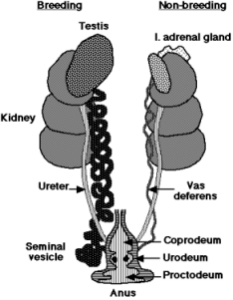
The Cloaca or vent has three parts: 1) the coprodeum that receives feces and
passes it to the 2) proctodeum the discharge area for eggs, and all waste and
the 3) Urodeum which recieves waste products from the kidneys and either eggs or
sperm and passes them to the proctodeum. In male ducks, swans and geese the
cloaca may form a copulatory organ.
The Reproductive Systems
The reproductive systems of birds have many many features that are similar to
reptiles including reduced size and weight that in birds helps enable flight,
seasonality and oviparity or reproduction by egg laying. Bird eggs have larger
yolks and porous shells that allow for the greater gas exchange needed by
warm-blooded animals.
The males have paired abdominal testes lying in front of the first kidney lobe.
The vas deferens runs from the testes to the cloaca where it has a common
opening with the ureter in the Urodeum. The terminal end of the vas deferens is
enlarged and acts as a storage organ called the seminal vesicle.
Copulation in birds involves meeting of the cloacal region of male and female
following courtship. This is often commonly referred to as cloacal kissing.
Female reproductive system
In most birds only the left ovary and oviduct are functional. This reduces
weight and makes only one developing egg needing to be supported at any given
time.
The ovary enlarges greatly during the breeding season and resemble small bunches
of grapes which are the developing egg follicles. The oviduct opens medially to
it in a funnel-shaped ostium - the equivalent of the mammalian infundibulum and
fimbriae.
The oviduct receives the developing eggs and has separate areas in which
different phases of egg formation occur. These are the magnum, the isthmus, the
uterus with pigment glands and the vagina for temporary egg storage.
Egg
formation.
Ovulation releases an egg from a mature follicle on the surface of the ovary.
The egg has food reserves in the concentric layers of the yolk. This is
immediately picked up by the ostium, where ciliary currents carry it into the
magnum region. Fertilization normally occurs here if the female has mated: sperm
are stored for extensive periods in glandular regions of the lower oviduct.
Over about three hours the egg receives a coating of albumen.
The egg and albumen then pass into the isthmus, where the shell membranes are
deposited. This takes about one hour.
In the uterine region the egg spends about a day, and pigment gets laid down in
the shell in characteristic patterns. Final hardening of the shell occurs here
and the egg then passes into the vagina and cloaca for laying.
![]()